XGT-9000

XGT-9000

X-ray Analytical Microscope (Micro-XRF)

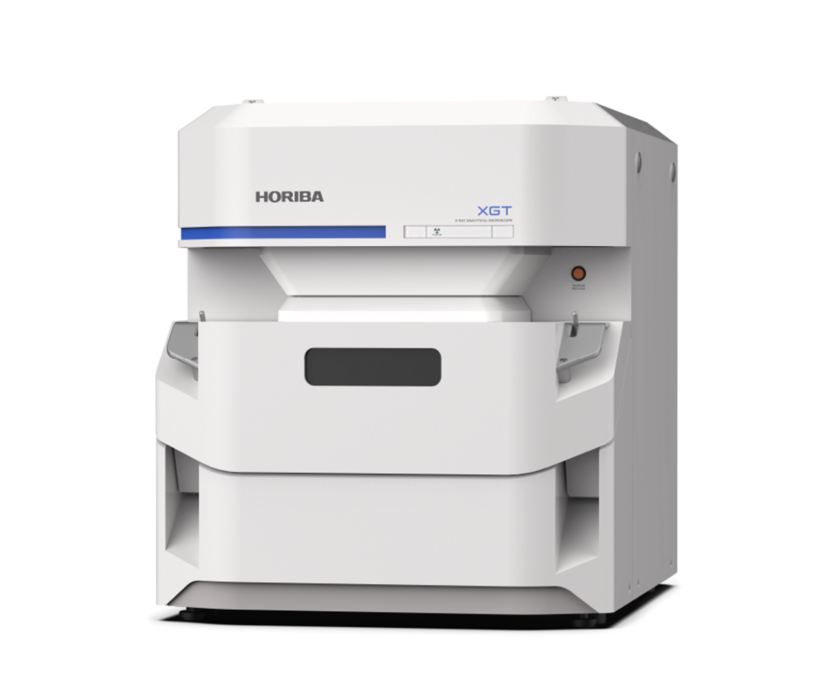
The XGT-9000 is a cutting-edge, non-destructive micro-XRF (X-ray fluorescence) analytical microscope offering exceptional speed, flexibility, and elemental mapping capabilities. It delivers ultra-fine spot analysis (down to ≤15 µm), dual-detector imaging (fluorescence and transmission), and multiple probe options—perfect for applications ranging from defect detection in electronics to archaeological artifact analysis.

Why Choose XGT-9000

  • Superior Sensitivity & Broad Element Range – Detects light elements through heavy metals with enhanced speed and precision.

  • Dual-Mode Imaging – Simultaneously captures fluorescence and transmission X-ray images for complete surface and internal structure insight.

  • Multi-Probe Flexibility – Offers fast scanning and high-resolution analysis by switching among a wide range of spot sizes.

  • User-Friendly Interface – Equipped with high-resolution cameras, versatile lighting, and intuitive software for streamlined operation.

Why Choose

Key Advantages of XGT-9000

Enhanced Detection Sensitivity

Ultra-Fast Analysis

Delivers high sensitivity across wide elemental ranges for rapid, accurate sample screening.

Comprehensive Imaging

Fluorescence + Transmission

Combines internal structure and surface elemental mapping in a single pass.

Adaptive Resolution

Selectable Probes

Switch easily between wide-area scanning and fine detail mapping using diverse spot sizes.

Intuitive Operation

Smart GUI & Lighting

User-friendly interface enhances workflow with clear imaging and customizable layouts.

Technical Specifications of XGT-9000


Model
XGT-9000
XGT-9000 ProXGT-9000 CXGT-9000 Expert


Basic information

   Instrument

X-ray analytical microscope
   PrincipleEnergy dispersive X-ray fluorescence spectroscopy
   Detectable elements*F (9) - Am (95)C (6) - Am (95)B (5) - Am (95)
   Available chamber size450 mm (W) x 500 mm (D) x 80 mm (H)
   Maximum mass of sample1 kg
   Maximum mapping area100 mm x 100 mm on 300 mm (W) x 250 mm (D)


Sample observation

   Optical image observationTwo high resolution cameras
   Whole image
   Detailed image
5 million pixels, Field of view: 100 mm x 100 mm
5 million pixels, Field of view: 2.5 mm x 2.5 mm
   Optical designVertical-coaxial X-ray and optical observation
   Sample illumination / ObservationTop, bottom, side illuminations / Bright and dark fields


X-ray generator

   PowerUp to 50 W
   VoltageUp to 50 kV
   CurrentUp to 1 mA
   Target materialRh


X-ray guide tube (Probe)

   Probe spot size selectionVarious probe combination can be offered
(e.g. 15μm ultra-high intensity probe with X-ray focused spot (Mo Kα))


Detectors

   X-ray fluorescence detectorLiquid nitrogen-free Silicon Drift Detector (SDD)
   Transmission detectorNaI (Tl)


Operating mode

   Measurement environmentWhole vacuum
Partial vacuum
Whole ambient
He purge (optional)
Whole vacuum
Partial vacuum
Whole ambient
He purge (optional)

Whole vacuum
Partial vacuum
He purge (optional)


Instrument dimension (main unit)

   Instrument size680 mm (W) x 860 mm (D) x 760 mm (H)
   Mass weightApproximately 200 kg
scrollable

*Under whole vacuum condition
 

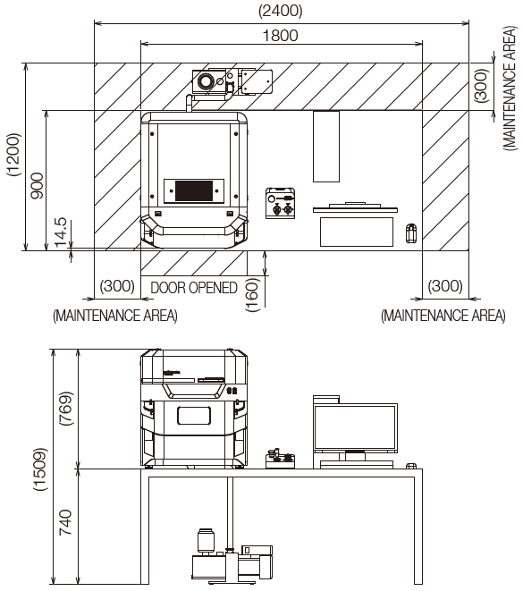
Dimensions (unit: mm) 
Related Products

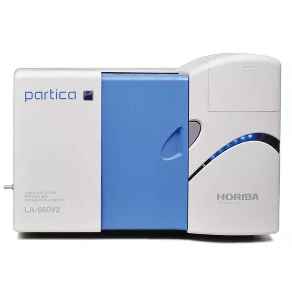
Partica LA-960V2

Partica LA-960V2

Laser Scattering Particle Size Analyzer

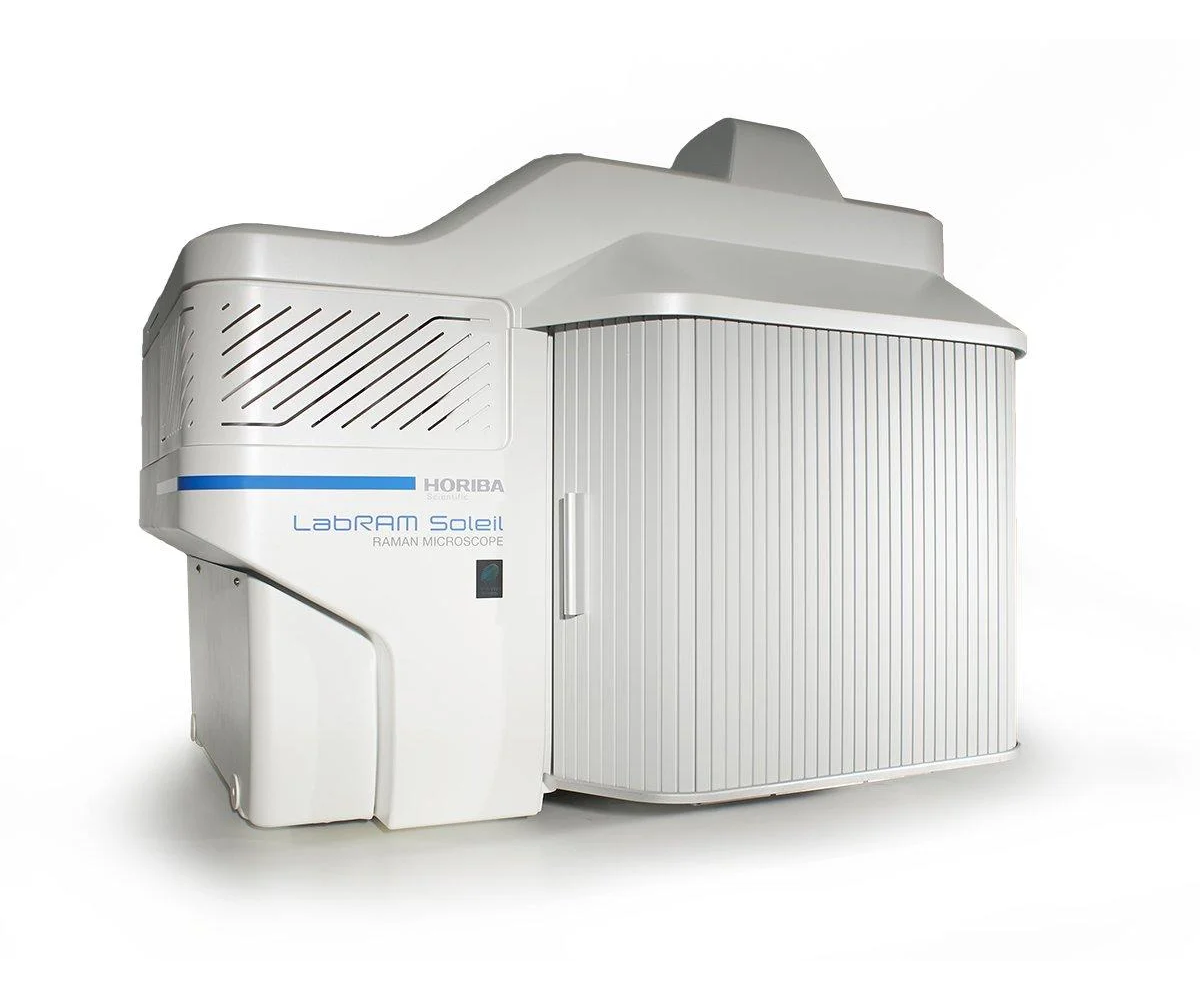
LabRAM Soleil

LabRAM Soleil

Confocal Raman Microscope with advanced automation and imaging features.

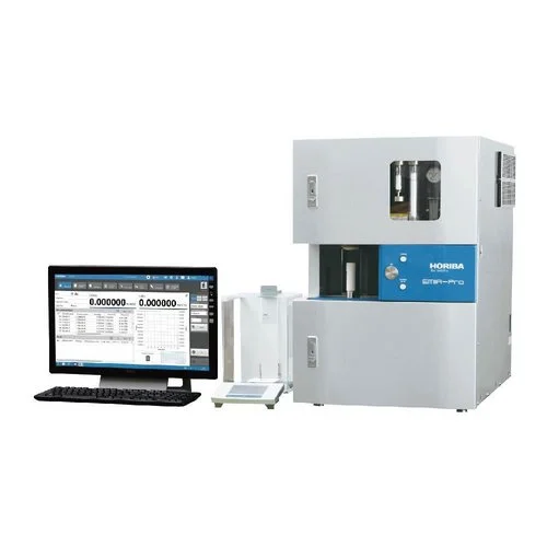
EMIA-Expert

EMIA-Expert

Carbon/Sulfur Analyzer (Flagship High-Accuracy Model)

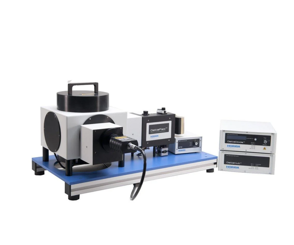
DeltaFlex

DeltaFlex

TCSPC Lifetime Fluorometer

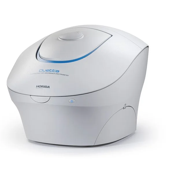
Duetta

Duetta

Fluorescence and Absorbance Spectrometer

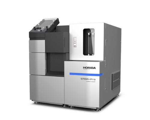
EMGA‑Pro (EMGA‑20P)

EMGA‑Pro (EMGA‑20P)

Oxygen/Nitrogen Analyzer

NOT SURE WHAT YOU ARE LOOKING FOR?

BOOK A CONSULTATION

Our experts are here to guide you to the right solution. Fill out the form and we'll get in touch shortly.

*
*
*
*
*