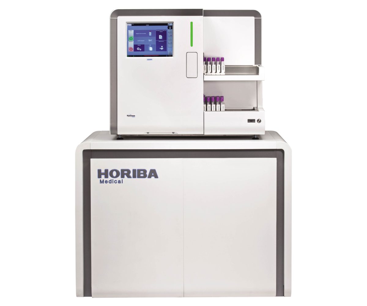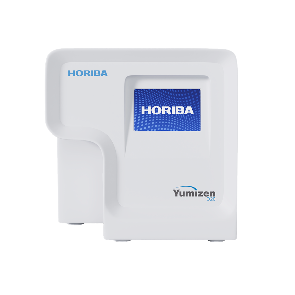
Yumizen D20
Digital Cell Morphology System
An in-vitro diagnostic device designed to automate manual microscopy in a diagnostic laboratory. It uses robotics and AI to digitize Blood and Urine samples to enable AI aided remote review. It generates a pre-classified report of blood and urine with accuracy. This report can be accessed and approved from anywhere, ensuring timely and accurate results to meet patient's needs.
The Yumizen D20 has 2 modules.
- Shonit: An AI application to analyze blood cell morphology which identifies and pre-classifies WBCs, RBCs and platelets in a peripheral blood smear.
- Shrava: An AI application to analyze urine sediment to identify and pre-classify multiple elements present in urine sediment.
Why Choose Yumizen D20
- Automate peripheral smear and Urine sedimentation reporting
- Accurate , Affordable and Accessible
- Cloud based platform for reporting of samples with unlimited storage.
- Seamless collaboration between doctors across geographies

Key Advantages of Yumizen D20
Dual Software
Offers two integrated platforms for peripheral smear and Urine samples for workflow management
High throughput
Processes 20 blood smear and 40 urine samples per hour reliably
CE Certified
Compatible with all staining protocol
Clinically proven
Continuously evolving algorithms ensure diagnostic accuracy
Applications of Yumizen D20
Complete Digital Hematology Solution
End‑to‑end automation with AI‑powered slide review, LIS integration, and real‑time reporting for fully digitized labs.
Government Hospitals & Medical College
Digitalization of Peripheral smear and Urine sample with centralized digital records for training and research.
Standalone Labs
Compact workflow solutions offering with AI and LIS integration with hematology analyzer & cloud reporting.
Corporate Hospitals + Level 5 Labs
Enterprise‑grade hematology AI based platform featuring multi‑site connectivity with standardized protocols and regulatory compliance management.
Technical Specifications of Yumizen D20
| Shonit module | Shrava module | |
| Analyzer type | Blood morphology analyzer | Urine sediment analyzer |
| Parameters | WBC – LYM%, MON%, NEU%, EOS%, BAS%, IG%, Atypical/Blasts%, NRBC%, Immature cells
RBC anisocytosis- Normocyte*, Microcyte*, Round Macrocyte*, Ovalo Macrocyte*
RBC Poikilocytosis – Elliptocyte*, Ovalocyte*, Target*, Teardrop*, Echinocyte*, Fragmented*
Platelets- PLT, Macro PLT, Giant PLT, Platelet Clumps *IHS Grades | Semi-Quantitative: RBC, PUS cells, Microorganisms
Detected vs Not Detected: Epithelial Cells, Cast, Crystal, Yeast
|
| Sample preparation | Manual or automated peripheral blood smears on glass slides with a thin layer of oil | Specially designed single-use cartridge GS200µ charged with 100µl of a uncentrifuged urine sample |
| Throughput | 20 slides per hour | 40 samples per hour |
| Slide/Cartridge loading | Single slide at a time | Single cartridge at a time |
| Slide/Cartridge specifications | Length – 74 to 76 mm Width – 24 to 26 mm Thickness – 1.0 to 1.35 mm | GS200µ cartridges |
| Oil immersion | A thin layer of oil to be applied manually | NA |
| Stains | Romanowsky Stains (Leishman, May-GrÜnwald- Giemsa, Wright Giemsa, Giemsa) | NA |
| Regulatory approval | CE IVDR | CE IVDR |
| Camera |
| |
| Optics system |
| |
| Illumination source | White LED | |
| Stage movement resolution |
| |
| Stage travel speed |
| |
| Barcode reader | External barcode reader | |
| Quality control |
| |
| Archiving of results and images | Unlimited archiving in Mandara utilizing either LAN or Wi-Fi | |
| External interface |
| |
| Electrical specifications |
| |
| Size (W x D x H) |
| |
| Weight |
| |
Related Products
NOT SURE WHAT YOU ARE LOOKING FOR?
BOOK A CONSULTATION
Our experts are here to guide you to the right solution. Fill out the form and we'll get in touch shortly.

